Alternative Splicing
Transcription introduced alternative splicing, where one gene joins its exons in more than one way to make several mRNAs, called isoforms, which can encode different versions of its protein. About 95% of human genes with more than one exon are alternatively spliced (Pan et al., 2008). This page looks at how the cell decides where to splice, the forms those decisions take, how they affect health and disease, and how to see them in sequencing data.
How Splice Sites Are Chosen
Splicing is done by the spliceosome, a large machine built from RNA and proteins. It recognizes short sequences at each end of an intron. The start of the intron is the 5′ splice site, or donor, and the end is the 3′ splice site, or acceptor. Nearly every intron starts with GT and ends with AG (GU and AG in the RNA). In a survey of mammalian splice sites, 98.71% used this pair, and 0.56% used GC and AG instead (Burset et al., 2000).
Two letters at each end can't be the whole story, since GT and AG turn up everywhere in the genome. Short stretches of sequence in exons and introns, called splicing enhancers and splicing silencers, are bound by RNA-binding proteins that make the spliceosome more or less likely to use a nearby site (Wang & Burge, 2008). Which of these proteins a cell makes, and how much of each, differs between cell types. Most alternative splicing events vary between tissues, so the same gene can be spliced one way in one tissue and another way in the next (Wang et al., 2008).
Types of Events
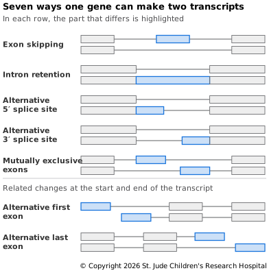
Figure 7.1 shows the main ways two isoforms can differ:
- Exon skipping. An exon is kept in one isoform and left out of the other.
- Intron retention. An intron stays in the mRNA. This affects up to three-quarters of mammalian genes with more than one exon, and it often lowers how much of the gene's mRNA there is (Braunschweig et al., 2014). One way is nonsense-mediated decay (NMD), a quality check that destroys mRNAs with a stop codon too early in them.
- Alternative 5′ splice site. The spliceosome uses a different donor, so the exon before the intron ends earlier or later.
- Alternative 3′ splice site. It uses a different acceptor, so the exon after the intron starts earlier or later.
- Mutually exclusive exons. Of two or more exons, exactly one is kept. The fruit fly gene Dscam takes this to the extreme, with alternative splicing that could make more than 38,000 isoforms (Schmucker et al., 2000).
The last two rows involve more than splicing. An alternative first exon depends on transcription starting at a different promoter. An alternative last exon depends on where the mRNA is cut and given its tail, called alternative polyadenylation. A different cut can also just lengthen or shorten the untranslated region in the same last exon. Wang et al. grouped these changes with the five splicing events, for eight types in all.
In every one of these events, the exons that are kept stay in the order they have in the gene. Most human pre-mRNAs are spliced into linear mRNAs that keep this order (Salzman et al., 2012), so an isoform can be exons 1, 2 and 4, with exon 3 skipped, but almost never 1, 4, 2 and 3. Transcripts of the gene DCC with exons joined out of order were found in 1991, at low levels and mostly without a poly-A tail (Nigro et al., 1991). Deep sequencing later showed that many such "scrambled" transcripts are circular RNAs, where the end of a later exon is joined back to the start of an earlier one (Salzman et al., 2012).
Why It Matters
Isoforms of the same gene can do different things, even opposite ones. BCL2L1 makes two well-studied proteins. The longer one, Bcl-xL, is 233 amino acids and protects cells from dying at least as well as its relative Bcl-2. The shorter one, Bcl-xS, is 170 amino acids and blocks Bcl-2 from protecting the cell. Bcl-xS was found at high levels in cells that turn over quickly, such as developing immune cells, and Bcl-xL in long-lived cells such as those of the adult brain (Boise et al., 1993; UniProt Q07817). The two isoforms differ by a single alternative 5′ splice site, which you'll find in Try It below.
When Splicing Goes Wrong
Because the spliceosome reads such short signals, small changes in DNA can redirect it. A variant that breaks a GT or AG can make the spliceosome skip an exon or use a nearby site instead. A variant elsewhere can create a new site that the spliceosome uses by mistake, called a cryptic splice site. These variants are easy to miss, because many lie in introns or don't change the amino acid they sit in (see Single-Nucleotide Variation and Insertions and Deletions). Using a deep learning model called SpliceAI, Jaganathan et al. estimated that 9% to 11% of disease-causing mutations in patients with rare genetic disorders act through cryptic splicing (Jaganathan et al., 2019).
Spinal muscular atrophy (SMA) shows what a difference one base can make. It's caused by the loss of both working copies of the gene SMN1, through deletion or mutation. A nearly identical gene, SMN2, codes for the same protein, but a C-to-T change in its exon 7, which doesn't change the amino acid, weakens a splicing enhancer and makes the spliceosome skip that exon in most of its mRNA (Lorson et al., 1999; Monani et al., 1999). One approved drug for SMA is an antisense oligonucleotide, a short synthetic strand that binds the SMN2 RNA in the intron after exon 7 and makes the spliceosome more likely to keep the exon (Hoy, 2017). In a trial in infants with SMA, 41% of those given the drug had a motor-milestone response at an interim analysis, compared with none in the control group (Finkel et al., 2017).
Splicing can also go wrong across many genes at once. In blood cancers with features of myelodysplasia, mutations in genes for parts of the splicing machinery, such as SF3B1, SRSF2, U2AF1 (U2AF35) and ZRSR2, were found in 45% to 85% of cases, depending on the type. Most of the mutated parts help recognize the 3′ splice site (Yoshida et al., 2011). For more on how cancer develops, see Cancer Biology.
Seeing It in Data
Sequencing reads show which exons were joined. In
RNA-Seq, a read
that crosses from one exon to the next shows that the two were spliced together, and a
splice-aware aligner such as STAR maps it in two pieces (see Read
Mapping). Each
short read covers at most a junction or two, so full isoforms have to be pieced
together. Long
reads
can cover a whole mRNA in one read, showing every exon of an isoform at once.
Annotations such as GENCODE list the known isoforms of each gene. In GTF/GFF3 Files, CFTR has 45 transcripts, and the MANE Select tag picks one to represent it. Which transcript you choose makes a difference when you describe a variant, since the same variant can fall in an exon of one isoform and an intron of another.
Setting Up
This page uses samtools and curl, as in Setting
Up in From Reads to Variants. If you
made the reads-to-variants or file-formats environment, activate it with conda activate. Otherwise, follow Install the
Tools on that page.
The files on this page take up about 200 MB.
Try It
Make a folder, then download the GENCODE annotation from GTF/GFF3 Files and pull out the lines for BCL2L1:
# Make a folder for this page and move into it
mkdir splicing && cd splicing
# Download the GTF file for GENCODE release 50
curl -fLO https://ftp.ebi.ac.uk/pub/databases/gencode/Gencode_human/release_50/gencode.v50.annotation.gtf.gz
# Save every line for BCL2L1 to a file
gunzip -c gencode.v50.annotation.gtf.gz | grep 'gene_name "BCL2L1";' > bcl2l1.gtf
# Show the first eight columns of the gene line
awk -F '\t' '$3 == "gene"' bcl2l1.gtf | cut -f 1-8
# Count its transcripts by type
awk -F '\t' '$3 == "transcript"' bcl2l1.gtf | grep -o 'transcript_type "[^"]*"' | sort | uniq -c | sort -rn
chr20	HAVANA	gene	31664452	31723989	.	-	.
  36 transcript_type "protein_coding"
   6 transcript_type "nonsense_mediated_decay"
   1 transcript_type "protein_coding_CDS_not_defined"
BCL2L1 is on chromosome 20, on the - strand, so the gene runs from high coordinates
to low. GENCODE lists 43 transcripts for it. Six are labeled nonsense_mediated_decay,
because their coding sequence ends more than 50 bases before a splice site, so NMD is
expected to destroy them (GENCODE
biotypes).
On the GTF page, you added up the CDS lines of one transcript to get the length of its
coding sequence. Do the same for every transcript at once, and divide by 3 to get each
protein's length in amino acids:
# Add up the CDS lengths of each transcript, divide by 3, and count the transcripts with each protein length
awk -F '\t' '$3 == "CDS" {split($9, a, ";"); n[a[2]] += $5 - $4 + 1} END {for (t in n) print n[t] / 3}' bcl2l1.gtf | sort | uniq -c | sort -rn
  17 233
   9 170
   6 200
   3 241
   2 307
   2 250
   1 251
   1 231
   1 207
split($9, a, ";") breaks the attributes apart at each semicolon, so a[2] is the
transcript ID, and n[a[2]] keeps a running total for each transcript. Many transcripts
make proteins of the same length. Seventeen code for 233 amino acids, the length of
Bcl-xL, and nine for 170, the length of Bcl-xS. Now compare one of each, BCL2L1-201
and BCL2L1-202:
# Save the lines for each transcript
grep 'transcript_name "BCL2L1-201";' bcl2l1.gtf > bcl2l1-201.gtf
grep 'transcript_name "BCL2L1-202";' bcl2l1.gtf > bcl2l1-202.gtf
# Show the protein length of each
awk -F '\t' '$3 == "CDS" {n += $5 - $4 + 1} END {print n / 3}' bcl2l1-201.gtf
awk -F '\t' '$3 == "CDS" {n += $5 - $4 + 1} END {print n / 3}' bcl2l1-202.gtf
# Show each exon's 5′ and 3′ ends, one row per exon, with BCL2L1-201 in columns 2–3
# and BCL2L1-202 in columns 4–5
paste <(awk -F '\t' '$3 == "exon" {print $5 "\t" $4}' bcl2l1-201.gtf) \
      <(awk -F '\t' '$3 == "exon" {print $5 "\t" $4}' bcl2l1-202.gtf) | awk '{print NR "\t" $0}'
233
170
1	31722868	31722618	31723529	31722618
2	31722348	31721655	31722348	31721844
3	31666086	31664458	31666086	31664452
BCL2L1-201 makes Bcl-xL and is the gene's MANE Select transcript, and BCL2L1-202
makes Bcl-xS. Both have three exons, and the GTF lists them in transcript order.
BCL2L1 is on the - strand, so it's read from high coordinates to low. Column 4 of a
GTF line is always the lower coordinate, whatever the strand, which means a - strand
exon begins at column 5 and ends at column 4. The command prints them in that order, so
each row reads from an exon's 5′ end to its 3′ end. Each transcript begins at the top
left of its pair of columns, at exon 1, and finishes at the bottom right, at the end of
exon 3.
Exon 1 differs only at its 5′ end, where transcription begins, and the protein doesn't start until exon 2. Exon 2 is where they part ways. Both copies begin at 31,722,348, but Bcl-xL's copy runs 31,721,844 − 31,721,655 = 189 bases further before it ends. That's an alternative 5′ splice site. The 189 bases are 63 codons, and 233 − 170 = 63. UniProt lists Bcl-xS as missing amino acids 126 to 188 of Bcl-xL. Exon 3 begins at the same place in both and ends 6 bases apart, after the stop codon.
To see the splice sites themselves, download chromosome 20 of the reference, as in From Reads to Variants:
# Download chromosome 20 of GRCh38 from UCSC
curl -fLO https://hgdownload.soe.ucsc.edu/goldenPath/hg38/chromosomes/chr20.fa.gz
# Decompress it to chr20.fa
gunzip chr20.fa.gz
# Index it so tools can jump to any position
samtools faidx chr20.fa
samtools faidx -i reads a region and reverse-complements it, so it shows the -
strand the way the gene reads it (samtools
faidx). The first two regions below are
the last 8 bases of exon 2 and the first 2 of the intron after it, for Bcl-xS and then
Bcl-xL. The third is the last 10 bases of the intron before exon 2:
# Read three splice sites on the - strand: the Bcl-xS donor, the Bcl-xL donor and the acceptor before exon 2
samtools faidx -i chr20.fa chr20:31721842-31721851 chr20:31721653-31721662 chr20:31722349-31722358
>chr20:31721842-31721851/rc
TTGAACAGGT
>chr20:31721653-31721662/rc
GCGGCTGGGT
>chr20:31722349-31722358/rc
TTTCCTGAAG
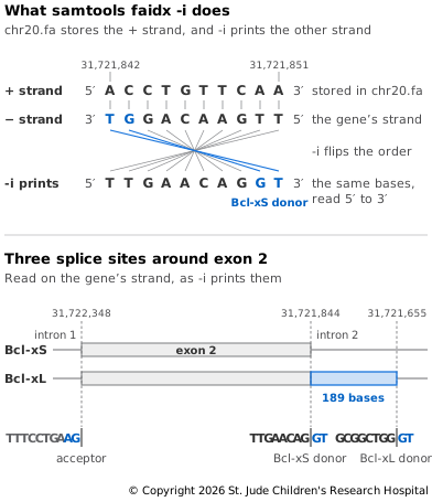
Figure 7.2 shows how -i gets these sequences and where they sit in the
gene. /rc marks each sequence as reverse-complemented. Both donors start the intron
with GT, 189 bases apart, and the acceptor ends it with AG. The spliceosome can use
either GT, and which one it picks decides whether the cell makes a protein that protects
it from dying or one that works against that protection.
That's the end of this exercise. Move out of the folder and delete it:
# Move up out of the folder
cd ..
# Delete the folder and everything in it
rm -r splicing