Translation
Decoding the Transcript
After DNA is transcribed to RNA and the RNA is spliced, the next step in protein production is translation. The genetic code that cells use to translate RNA to protein is nearly universal. You, your dog, and the lettuce in your salad all read it the same way; only mitochondria and a few microbes use slight variants. Because of this, a human gene placed in bacteria still produces the human protein, which is how insulin for diabetes is made. A shared code, together with the many genes humans share with other animals, is also why studying a gene in mice can tell us about the same gene in people.
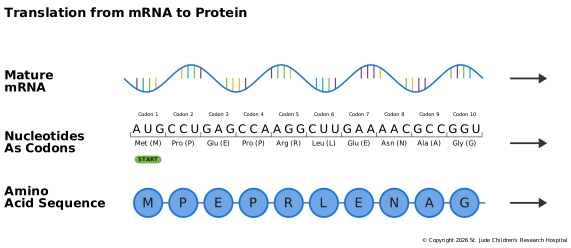
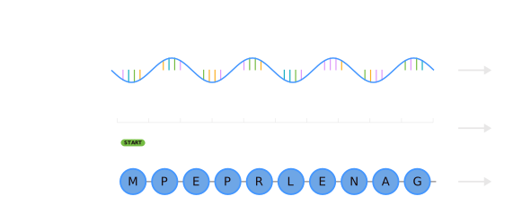
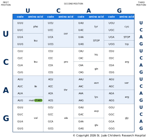
The mRNA transcript is encoded in three-letter nucleotide blocks known as codons. Codons specify that a particular protein building block, or amino acid, should be added to a growing protein chain. For example, the mRNA sequence above has "CCU" in the second codon position. This codon instructs the protein assembling machinery of the cell to add a proline to the amino acid sequence at position two.
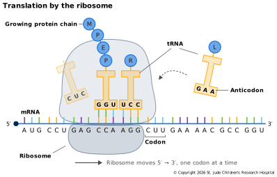
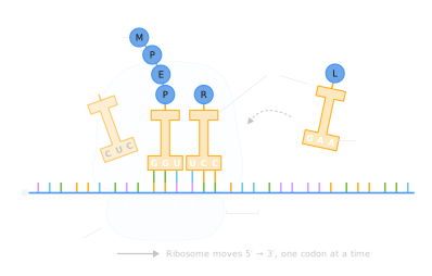
A ribosome moves along the mRNA one codon at a time, from the 5′ end toward the 3′ end. For each codon, a small adapter molecule called a transfer RNA (tRNA) brings in the matching amino acid: one end of the tRNA carries the amino acid, and the other end has three bases, called an anticodon, that pair with the codon. The ribosome joins each new amino acid to the growing chain until it reaches a stop codon, then releases the finished protein.
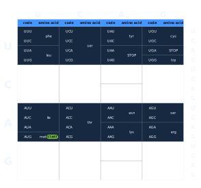
As shown in the amino acid lookup table below, other codons specify that translation should start or stop at a particular position. Because multiple codons translate to the same amino acid, some changes to the nucleotide sequence may not change which amino acid is added to the growing protein chain (e.g., if your triplet starts with "CG", the amino acid is always an arginine and the third nucleotide doesn't matter).
A change that alters the mRNA sequence but not the resulting protein is called a synonymous variant, often called a "silent mutation". Synonymous variants are usually harmless, though occasionally they affect splicing or how quickly a protein is made. Other changes can affect the final protein in ways ranging from insignificant to harmful. Knowledge of the codons and the chemistry of proteins is used to predict the effect of a given variant on the resulting protein.
Reading Frames
Whenever you decode one alphabet into another, accuracy depends on starting in the right place. Any stretch of mRNA can be split into codons in three different ways, called reading frames, depending on whether you start at the first, second or third nucleotide, and each frame spells out a completely different string of amino acids. The ribosome sets the frame by scanning along the mRNA from the 5′ cap until it finds a start codon, AUG (which codes for methionine), usually the first one it meets. From there it reads three bases at a time.
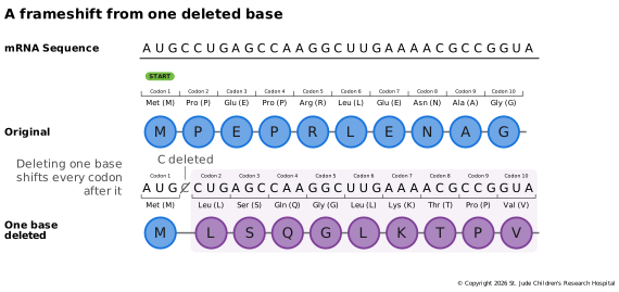
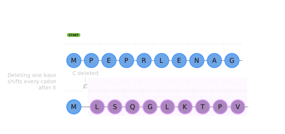
Consider the sequence in the figure above (a contrived example, but useful for our illustration here). Read from its start codon, it gives methionine, proline, glutamic acid and so on. Now suppose a single nucleotide, the C just after the start codon, is deleted. The ribosome still starts at AUG, but every codon after the deletion is read one position further along: "CUG" (leucine) instead of "CCU" (proline), and a completely different protein is made from that point on.
An open reading frame (ORF) begins with a start codon and runs to a stop codon, with no other stop codon in between. This is the stretch that is translated into a protein. Changes that create or remove a start or stop codon, or that shift the reading frame, can dramatically alter the protein. An insertion or deletion whose length is not a multiple of three shifts every codon after it, as in the example above; this is called a frameshift mutation.
Bakery Analogy
On the baking floor, the baker (a ribosome) reads the index card (mRNA) in three-letter words (codons). The baker starts at the word that marks the beginning of the recipe (the start codon) and stops at the word that marks its end (a stop codon). For each word, a kitchen assistant (a tRNA) brings the matching ingredient (an amino acid) from the pantry. A typo that turns one word into another word for the same ingredient makes no difference to the cake (a synonymous variant). But if a letter is dropped, every word after it is misread (a frameshift), and the cake comes out completely different.