Baselines for Variation
A variant is a difference, which raises an awkward question: different from what? There is no single "normal" human genome to compare against. Any two people differ at millions of positions, and as the Introduction explained, even the cells within one body slowly drift apart. Genomics settles the question practically: everyone agrees to compare against the same sequence.
The Reference Genome
That shared sequence is the reference genome. In practice it is a file of about 3.1 billion bases, with one long sequence for each chromosome. You have two copies of each, but the reference has just one, so it can't show both of a person's haplotypes. Nor is it any one person. About 70% of it comes from a single anonymous volunteer from Buffalo, New York, known as RP11, and the rest is stitched together from more than 20 others.
Because everyone uses the same reference, any position in the genome can be named by a
coordinate: the chromosome, then the position counting from its start. F508del, for
example, sits at chr7:117,559,592. At each position, the base in the reference is
called the reference allele (often shortened to REF), and a different base seen in a
sample is an alternate allele (ALT). These are the names of two columns you will
meet in VCF files.
The reference also gives genotypes a fixed point of comparison. You have two copies of
most positions, so the labels homozygous and
heterozygous
can be stated relative to it. If both copies carry the reference allele, the genotype is
homozygous (ref). If one carries the reference allele and the other an alternate
allele, it is heterozygous. If both carry the same alternate allele, it is homozygous
(alt). Nearly every position in anyone's genome is homozygous (ref), so a list of
variant calls usually includes only the other two. VCF files write these three as 0/0,
0/1 and 1/1. Each number stands for the allele on one of the two copies: 0 is the
reference allele and 1 is the alternate allele. If a position has a second alternate
allele, it is written as 2.
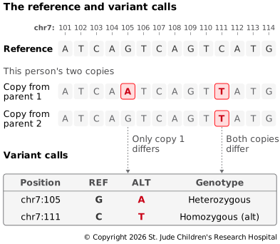
The reference allele only means "whatever the reference has". It isn't necessarily the healthy version or even the most common one. At many positions the reference carries the rarer allele. Historically, it's even carried alleles linked to a higher risk of disease, including type 1 diabetes.
Versions and Their Traps
The human reference is maintained by the Genome Reference Consortium (GRC), which also
looks after the mouse, zebrafish and chicken genomes. The current version is GRCh38:
GRC for the consortium, h for human and 38 for the version. The UCSC Genome
Browser calls the same version hg38, and the previous version, GRCh37, is known
there as hg19. The GRC also releases patches (the latest is GRCh38.p14, from
2022), which fix errors and add sequence without moving any existing coordinates.
Versions cause some of the most common mistakes in genomics:
- Coordinates don't carry over between versions. F508del is at
chr7:117,559,592in GRCh38 butchr7:117,199,646in GRCh37. Always record which version your data uses. Converting positions from one version to another is called liftover, and it can fail in regions that changed a lot. - Chromosome names differ. UCSC names chromosomes
chr1,chr2andchrM, while the GRC and Ensembl use1,2andMT. Tools given files with different conventions often report no matches at all instead of raising an error. - Similar names hide small differences.
hg19andGRCh37have the same chromosome sequences, but different mitochondrial sequences, and distributions such asGRCh37-liteadd or leave out extra sequences. Files built on one don't always work with another. If you have to work with older versions, the Wikipedia page has a helpful table comparing them.
How Common Is a Variant?
Comparing against the reference says only that a sample is different. To learn whether that difference is unusual, analysts turn to population frequency databases such as gnomAD, which collects sequencing data from hundreds of thousands of people and records how often each allele appears, overall and within different ancestry groups. A variant carried by one person in ten is very unlikely to cause a rare, severe disease, while one never seen before deserves a closer look (as does one seen frequently in people with the disease). That last kind of evidence comes from a different source: databases such as ClinVar collect reports from labs and researchers on which variants have been linked to disease, and how confidently.
Beyond a Single Reference
One genome, mostly from one person, can't represent everyone. Because so much of the reference is RP11, his DNA quietly became the yardstick for everyone else's. Wherever he happened to carry a rare allele, most people show up as "different", and any stretch of DNA he lacked can be missing from the reference altogether. A 2019 study of 910 people of African descent found about 300 million bases of DNA missing from the reference. Reads from those regions have nowhere to map, so the variants in them are easy to miss, and people whose ancestry is less well represented are affected most.
Two newer efforts are filling the gaps. In 2022, the Telomere-to-Telomere Consortium published T2T-CHM13, the first complete, gap-free human genome, adding about 8% of the genome that GRCh38 was missing. In 2023, the Human Pangenome Reference Consortium released a draft human pangenome built from 47 people, and a second release in 2025 expanded it to 232. Instead of one sequence, a pangenome stores many genomes together, branching wherever they differ. That eases the yardstick problem, but a graph brings challenges of its own:
- No single set of coordinates. A position in a graph depends on which path you take through it, while gene annotations, disease databases and VCF files all use linear coordinates. Today's pangenome workflows usually map reads to the graph, then translate the results back onto GRCh38.
- No single right way to build one. Aligning many whole genomes involves judgment calls, especially for repeated and duplicated DNA. In the draft pangenome, one construction method merged the extra copies of duplicated genes and repeats into a single copy, while the others kept them separate. Another left out most centromeric repeats, which are hard to align and often misassembled.
- More variation can make mapping harder. Every rare branch is another place a read might plausibly land. The draft pangenome's graph for mapping short reads leaves out any sequence carried by fewer than 9 of its 90 genome copies, about 10%.
- New tools and formats. Graphs need their own file formats and software, while most of the ecosystem, including the rest of this guide, is still built around a linear reference.
- Only as diverse as the people in it. A few hundred people is still a small sample of humanity, and any group left out faces the same gaps as before.
In the meantime, the GRC has postponed GRCh39 indefinitely while it evaluates these new models, and most data is still analyzed against GRCh38.
A Second Baseline: The Person's Own DNA
For somatic variants, the best baseline is the person themselves. In cancer, analysts often sequence the tumor alongside a matched normal sample, usually blood, from the same person. Variants found in both are germline, and variants found only in the tumor are likely somatic. Both are important: for example, an inherited variant in BRCA1 raises the risk of breast and ovarian cancer, while the somatic variants are what drive this particular tumor.
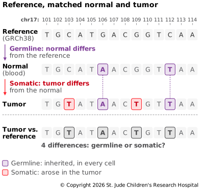
Without a matched normal, a tumor-only analysis has to guess which variants are germline, usually by checking how common they are in population databases. That works less well, especially for people whose ancestry is underrepresented in those databases. Studies of germline samples alone answer a different question: which variants run in families or raise the risk of disease.