Single Nucleotide Variation
A single nucleotide variant (SNV) is the smallest change a genome can carry: one base swapped for another at a single position. Most of the four to five million differences in a typical genome (see the Introduction) are SNVs.
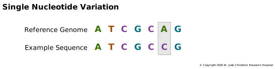

One change like this can have a large effect. In many melanomas, the tumor has a T at
chr7:140,753,336 (GRCh38) where the reference has an A. That single change, known as
BRAF V600E, swaps one amino acid in the BRAF protein and leaves it permanently
switched on, pushing the cell to keep dividing.
SNVs and SNPs
A single nucleotide polymorphism (SNP, pronounced "snip") is an SNV that is common in a population, usually defined as carried by more than 1% of people, though there is no agreed cutoff. Every SNP is an SNV, but not every SNV is a SNP. In practice the line blurs: people say "snips" for any SNV, since "SNV" has no easy pronunciation, and the dbSNP catalog, despite its name, includes rare variants and even small insertions and deletions.
In cancer, the more useful split is germline versus somatic. A variant that is common in the population is almost always inherited, which is why tumor-only analyses use frequency databases to set common variants aside. Common SNPs can still be important: some raise or lower cancer risk, and others change how the body handles drugs.
What an SNV Can Do
The effect of an SNV depends on where it lands. Most fall outside genes, or in parts of genes that don't encode protein, and have little or no effect. Inside a coding sequence, the codon table predicts the result:
- Synonymous. The new codon encodes the same amino acid, so the protein is unchanged.
- Missense. The new codon encodes a different amino acid, as in BRAF V600E. A missense variant can be harmless or severe, depending on which amino acid changes and where it sits in the folded protein.
- Nonsense. The new codon is a stop codon, cutting the protein short. A nonsense variant is what G542X is in CFTR.
SNVs outside the coding sequence can also have effects. One at a splice site can make the cell skip an exon or keep an intron, and one in a regulatory region can change how much of a gene is made.
Where Somatic SNVs Come From
Where a somatic SNV lands is largely a matter of chance, but which change it makes often isn't. Every cell picks up a slow, steady trickle of SNVs from copying errors and ordinary chemical wear, such as the gradual decay that turns some Cs into Ts, so these build up with age. Outside sources of damage add their own: ultraviolet light mostly turns C into T, often two neighboring Cs at once (CC to TT), and tobacco smoke favors C to A. The mix of SNVs in a tumor therefore records what caused them. These patterns are called mutational signatures, and the COSMIC signature catalog helps analysts match a tumor's pattern to its likely causes.
Because each process leaves its own pattern, software can now work backward from a tumor to its history. Tools such as SigProfiler and deconstructSigs sort a tumor's SNVs into 96 categories (six kinds of base change, each split by the bases on either side) and estimate how much each known signature contributed. The answer can point outside the body, to sunlight or smoking, or to problems inside the cell. A tumor that can't repair its DNA properly, for example because of an inherited BRCA1 variant, leaves a recognizable signature. Some chemotherapy drugs leave a mark too, which shows up in tumors that return after treatment. These are statistical estimates, though, and they need plenty of mutations to work with; a tumor with only a few dozen SNVs gives them little to go on.
Linking SNVs to Disease
A tumor can carry anywhere from a handful to hundreds of thousands of somatic SNVs, and a germline genome carries millions. Each one tells you something, as mutational signatures show, but in cancer the pressing question is which few actually raise the risk of the disease or drive its growth.
One approach is statistical. A genome-wide association study (GWAS) compares people with a disease (cases) to people without it (controls), looking for variants, usually common SNPs, that turn up in cases more often than chance would explain. A hit rarely pinpoints the cause on its own. Nearby variants are inherited together on the same haplotype, so the SNP that stands out may only mark a region, and the variant that actually causes the effect may be one of its neighbors. Most common diseases also involve many variants, each with a small effect. Because GWAS needs thousands of samples, rare diseases are harder to study this way, but not impossible: in 2009, two studies found inherited variants in ARID5B and IKZF1 that raise the risk of childhood acute lymphoblastic leukemia.
For the somatic SNVs in a single tumor, analysts lean on biology instead. They look closely at SNVs that change a protein, especially in genes already known to be involved in cancer, and check every candidate against databases such as COSMIC, which catalogs somatic variants found in tumors. The strongest clue is recurrence: a variant that turns up at the same position in many tumors, as BRAF V600E does, is likely a driver that helps the cancer grow. Most of the rest are passengers, changes that came along for the ride.
Recurrence counts outside genes too. In about 70% of melanomas, and in several
other cancers, one of two SNVs sits in the
promoter of TERT. Each creates a
new binding site for transcription factors and turns up production of telomerase, an
enzyme that lets cells keep dividing indefinitely. The more common one, at
chr5:1,295,113 in GRCh38, is known as C228T after its GRCh37 position,
chr5:1,295,228, a reminder that coordinates depend on the version. Drivers like these
are easy to miss: exome sequencing and many gene panels read only the coding parts of
genes, so finding them takes whole-genome sequencing or a panel designed to include
them.
Figure 3.4 shows what recurrence looks like in real data. It plots the somatic BRAF variants found in tumors sequenced for the NCI Genomic Data Commons along the length of the protein.
Loading BRAF variants from ProteinPaint…
A lollipop plot of the BRAF protein, about 770 amino acids long, drawn as a bar with its domains shaded. Somatic variants from tumors stand above it as circles on stems, sized by how many tumors carry each one. Small circles are scattered along the whole protein, smaller clusters sit at positions 466, 469, 581 and 594, and one circle at position 600, V600E, is far larger than all the others: it is found in several hundred tumors.
Somatic variants in BRAF from tumors in the NCI Genomic Data Commons. Each circle is one protein change, sized by the number of tumors that carry it, and its color shows the kind of change (blue for missense). V600E dwarfs everything else. Drawn with ProteinPaint (Zhou et al., 2016), which loads current data, so the counts grow as new tumors are added.