The Structure of DNA
The main text treats DNA as a long string of A, C, G and T. That abstraction is all you need for most genomics work, but a few everyday terms, such as 5′ and 3′, the template strand, and the reverse complement, only make sense once you look at how the molecule is built. This guide covers those details.
Nucleotides
DNA is a chain of building blocks called nucleotides. Each nucleotide has three parts:
- a phosphate group;
- a five-carbon sugar called deoxyribose;
- one of four bases: adenine (A), guanine (G), cytosine (C) or thymine (T).
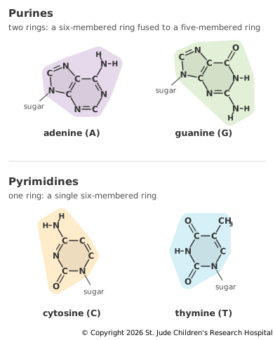
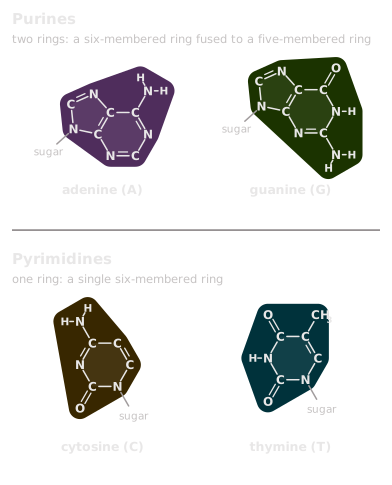
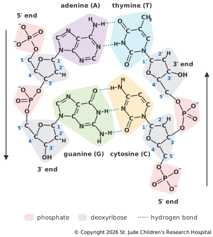
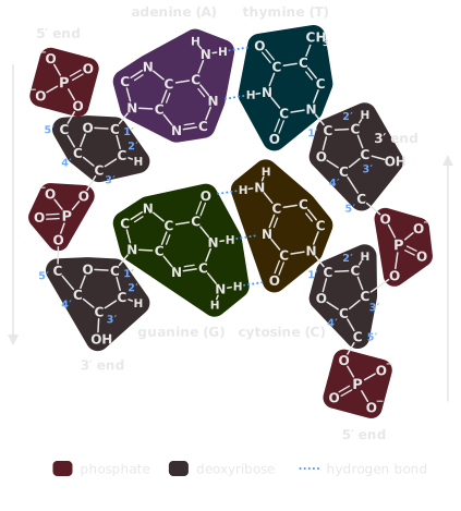
A and G are purines, which are larger and have two rings: a six-membered ring of carbon and nitrogen atoms fused to a five-membered ring. C and T are pyrimidines, which are smaller and have a single six-membered ring. Within each group, the bases share the same ring framework and differ in the atoms attached to it. Adenine carries an NH2 (amino) group, and guanine carries an oxygen and an NH2 group. Cytosine carries an NH2 group and an oxygen, and thymine carries two oxygens and a CH3 (methyl) group. Several of these attached atoms, along with nitrogens in the rings, form the hydrogen bonds that hold base pairs together (Figure 7.10).
The "deoxy" in deoxyribonucleic acid means the sugar is missing one oxygen atom that the sugar in RNA, ribose, still has.
The Backbone and Its Direction
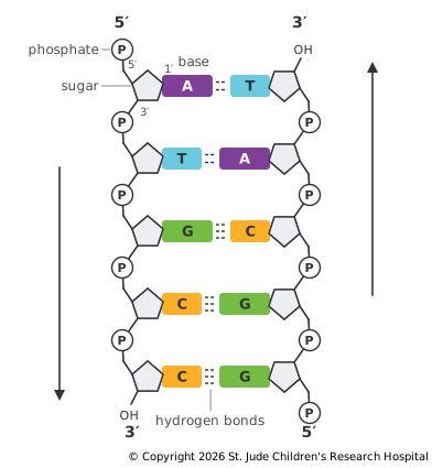
The five carbon atoms in each sugar are numbered 1′ to 5′ ("one prime" to "five prime"). The prime mark keeps them distinct from the numbered atoms in the base. The base is attached to the 1′ carbon, and the oxygen that deoxyribose lacks is the one on the 2′ carbon.
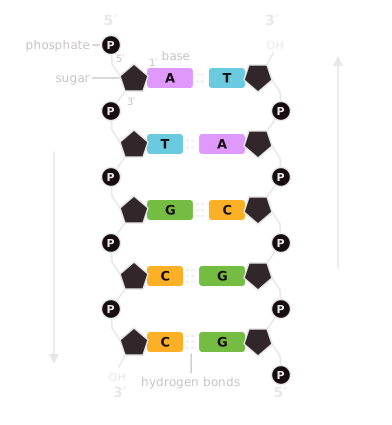
Nucleotides join into a strand through their phosphates: each phosphate links the 3′ carbon of one sugar to the 5′ carbon of the next. This repeating sugar-phosphate chain forms the backbone of the strand, and the bases stick out from it to one side.
Because every link runs from a 3′ carbon to a 5′ carbon, a strand has a direction. One end, the 5′ end, finishes with a 5′ carbon (usually carrying a phosphate), and the other, the 3′ end, finishes with a 3′ carbon. By convention, sequences are always written from 5′ to 3′. Figure 7.9 shows the backbone and both ends of each strand, and Figure 7.10, in the next section, numbers the carbons of every sugar.
The direction matters because the enzymes that build DNA and RNA can only add new nucleotides to the 3′ end of a growing strand, so every new strand is built from 5′ to 3′. For mRNA, this means:
- the front of the mRNA, made first, is its 5′ end, which is where the 5′ cap goes;
- the back of the mRNA, made last, is its 3′ end, which is where the poly-A tail goes;
- ribosomes also read mRNA from 5′ to 3′, so they start at the cap end.
Two Strands, Opposite Directions
The two strands of a DNA double helix run in opposite directions, which is called antiparallel. Where one strand runs 5′ to 3′ from left to right, its partner runs 5′ to 3′ from right to left.
The strands are held together by hydrogen bonds between paired bases. A pairs with T through two hydrogen bonds, and G pairs with C through three. GC-rich DNA is harder to pull apart. The extra hydrogen bond explains only part of this. About half of the difference comes from G and C stacking more strongly on the bases next to them (Yakovchuk et al., 2006). Every pair joins a larger purine to a smaller pyrimidine, so each rung of the ladder in Figure 7.4 is the same width.
The paired strands twist into a right-handed helix about 2 nanometers wide (Watson and Crick, 1953). In solution, each full turn spans about 10.4 base pairs (Wang, 1979). The twist leaves two grooves of different sizes, the major groove and the minor groove, along the outside of the helix. Proteins such as transcription factors read the edges of the bases exposed in these grooves, so they can recognize a sequence while the DNA stays double-stranded (Seeman et al., 1976; Rohs et al., 2010).
Reading the Other Strand
Because the strands are antiparallel and their bases pair, each strand determines the other. To write the partner of a sequence from 5′ to 3′, reverse it and swap each base for its partner. The result is called the reverse complement:
Strand:              5′-ATGCC-3′
Partner in helix:    3′-TACGG-5′
Reverse complement:  5′-GGCAT-3′
This explains the template and coding strands from Transcription. RNA polymerase reads the template strand from 3′ to 5′ so that it can build the RNA from 5′ to 3′. The RNA is therefore the reverse complement of the template strand, which makes it the same sequence as the coding strand, with U in place of T.
The reverse complement also comes up constantly in sequencing data. A sequencer reads whichever strand a DNA fragment came from, so a read may match the reference genome directly or only after taking its reverse complement. Aligners check both, and file formats record which strand each read matched.
How RNA Differs
RNA uses the same backbone chemistry and the same 5′ and 3′ ends, with three differences:
- its sugar is ribose rather than deoxyribose;
- it uses uracil (U) in place of thymine (T);
- it is usually single-stranded, and often folds back on itself where short stretches of its own sequence pair up.
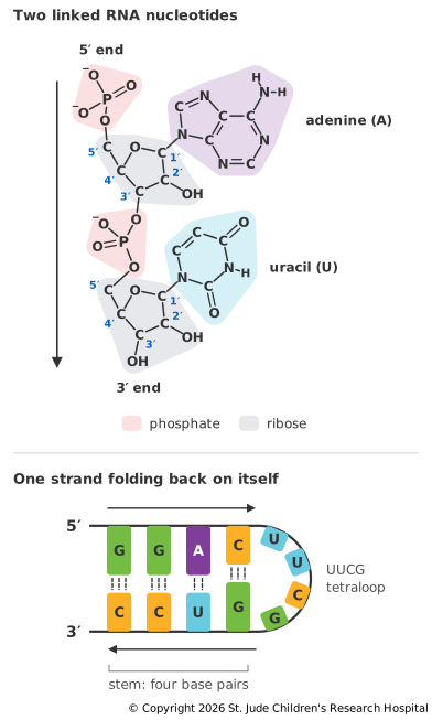
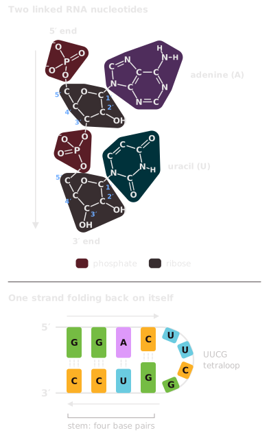
The extra oxygen in ribose, part of an OH group on the 2′ carbon, makes RNA less chemically stable than DNA. It can attack the neighboring phosphate and break the backbone (Soukup and Breaker, 1999). This suits their roles: DNA is the long-term record, and mRNAs are short-lived working copies. Other RNAs, such as those that make up ribosomes, are far more stable.