Epigenetic Modifications
Nearly every cell in your body carries the same genome, yet a neuron and a white blood cell look and behave nothing alike. What differs is which genes each cell uses. Part of what holds those choices in place is a layer of chemical marks on the DNA and on the histones it wraps around. Epigenetics is the study of changes in gene activity that can be passed on when a cell divides but can't be explained by changes in the DNA sequence (Bird, 2002). This guide covers the two best-studied kinds of marks, how they're measured and what happens to them in cancer.
DNA Methylation
The most common mark on human DNA is a methyl group, one carbon atom with three hydrogens (CH3), attached to carbon 5 of a cytosine. The result is called 5-methylcytosine (Moore et al., 2013). In most human cells it's found almost only at CpG sites, where a C is followed by a G on the same strand; the "p" stands for the phosphate that links them. CG read 5′ to 3′ is its own reverse complement, so the other strand has a CpG at the same spot, and the two cytosines sit diagonally across from each other (Figure 7.3). In human body cells, 70% to 80% of all CpG sites are methylated, as Bird's review notes.
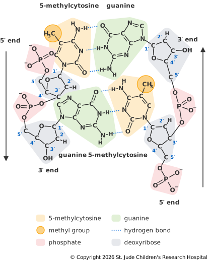
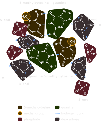
CpG sites are scarce across most of the genome but cluster in stretches about 1,000 bases long called CpG islands, which normally stay unmethylated (Deaton and Bird, 2011). About 70% of gene promoters sit in a CpG island (Saxonov et al., 2006). Dense methylation of a promoter's island helps lock the gene in a silent state. Bird's review names two places where cells do this on purpose during development: the inactive X chromosome in females, and imprinted genes, where only the copy inherited from one parent is used.
Copying the Marks
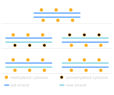
When a cell divides, its methylation is copied along with its DNA, so the marks outlast the cell that made them (Figure 7.4). A new strand is built from plain cytosines, so right after copying, each CpG that was methylated on both strands now carries a methyl group on the old strand only, a state called hemimethylated. A protein called UHRF1 binds these half-marked sites (Bostick et al., 2007), and the enzyme DNMT1 adds a methyl group to the new strand's C wherever the old strand has one. The copying isn't perfect, so now and then a site loses its mark.
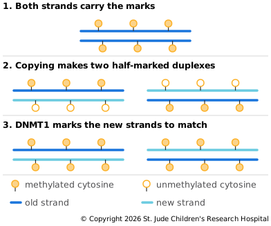
Other enzymes change the pattern. DNMT3A and DNMT3B add methyl groups to sites that had none (Okano et al., 1999). TET enzymes start the work of removing marks by converting 5-methylcytosine to 5-hydroxymethylcytosine (Tahiliani et al., 2009).
Histone Modifications
Histones are the proteins DNA wraps around. Each nucleosome is a core of eight histones with 145 to 147 base pairs of DNA wound around it, and flexible tails, 15 to 36 amino acids long, stick out from it (McGinty and Tan, 2015). Enzymes attach small chemical groups to particular amino acids in these tails, most often lysines.
This page covers two kinds of marks (Bannister and Kouzarides, 2011):
- Acetylation adds an acetyl group to a lysine. That cancels the lysine's positive charge and can weaken its hold on DNA. Enzymes called histone acetyltransferases add these marks, and histone deacetylases remove them.
- Methylation adds one, two or three methyl groups to a lysine. The same lysine can carry either kind of mark.
Each mark is named with a short code: the histone, then the amino acid and its position in the tail, then the mark (Figure 7.5). H3K27me3 means three methyl groups (me3) on lysine (K) 27 of histone H3, and H3K27ac is an acetyl group on the same lysine. Positions are counted without the methionine that starts the protein, because the cell trims it off soon after the protein is made (Blasco-Santana et al., 2024).
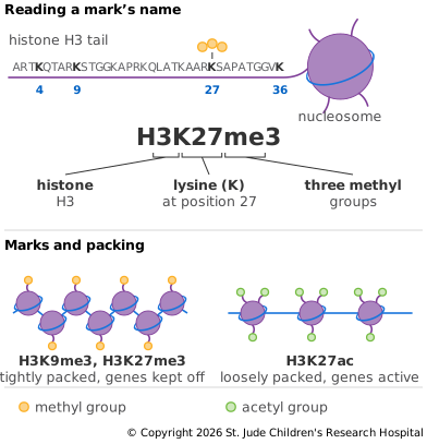
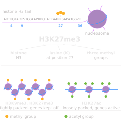
Each mark is associated with a particular kind of region. The Roadmap Epigenomics project mapped the most common ones in 111 human tissues and cell types (Roadmap Epigenomics Consortium, 2015):
| Mark | Usually found at |
|---|---|
| H3K4me3 | Promoters |
| H3K4me1 | Enhancers |
| H3K27ac | Active promoters and enhancers |
| H3K36me3 | The bodies of genes being transcribed |
| H3K27me3 | Genes switched off by Polycomb proteins, which may be on in other cells |
| H3K9me3 | Heterochromatin, especially repeat-rich DNA that is usually packed tight |
H3K27me3 is made by PRC2, a group of Polycomb proteins that helps cells keep their identity (Margueron and Reinberg, 2011). It usually marks DNA that's packed away in one cell type but may be opened in another, while H3K9me3 tends to mark DNA that stays packed across many cell types (Saksouk et al., 2015).
These tendencies have exceptions. In embryonic stem cells, many developmental genes carry H3K4me3 and H3K27me3 at once, which keeps them off but ready to switch on later (Bernstein et al., 2006). A family of active genes that code for zinc finger proteins carries H3K9me3 near their ends (Blahnik et al., 2011). And when engineered cells lost most of the H3K27ac at their enhancers, their genes' activity barely changed (Zhang et al., 2020). A mark tells you what usually goes on at a region, but it doesn't always cause it.
Some histone marks can also be passed on when a cell divides, at least those that keep genes off. When DNA is copied, the old histones are shared between the two new copies, so each inherits about half the marks, and enzymes that recognize a mark and add more of it fill in the rest. For H3K9me3 and H3K27me3, that refilling can take more than one cell cycle (Alabert et al., 2015). In fruit flies, H3K27me3 carried a gene's off state through several divisions (Coleman and Struhl, 2017), though keeping it going also needed the DNA sequences that call in PRC2 (Laprell et al., 2017). Histones from active regions aren't kept in place the same way (Escobar et al., 2019), so whether marks of active genes are inherited is less clear.
Measuring the Marks
Standard sequencing reads 5-methylcytosine as an ordinary C, and PCR copies lose the marks, so methylation needs its own methods.
- Bisulfite sequencing treats DNA with sodium bisulfite, which turns unmethylated cytosine into uracil but leaves 5-methylcytosine alone. PCR then copies each uracil as T (Frommer et al., 1992). After alignment, a C that stayed C was methylated, and a C that became T wasn't. Run across a whole genome, it maps methylation at nearly every C. One such study found that 99.98% of the methylation in a fibroblast cell line was at CpG sites, while in embryonic stem cells nearly a quarter was elsewhere (Lister et al., 2009). Bisulfite can't tell 5-methylcytosine from 5-hydroxymethylcytosine, because neither one becomes uracil, so both are read as C (Huang et al., 2010).
- Enzymatic methyl-seq (EM-seq) makes the same distinction with enzymes in place of bisulfite, which damages and breaks DNA. Because it's gentler, it works with as little as 100 picograms of DNA (Vaisvila et al., 2021). Like bisulfite, it reads both modified forms as C.
- Long reads come from the original molecule, so methylation changes the signal itself and can be detected without any conversion.
Histone marks are measured through the DNA they're attached to. ChIP-seq uses an antibody against one mark, such as H3K27me3, to pull out the DNA wrapped around nucleosomes carrying it. ATAC-seq shows which stretches of DNA are open enough for an enzyme to reach, though it can't say which marks are present.
Epigenetics in Cancer
Too Little and Too Much Methylation
In 1983, two labs independently reported that cancer cells carry less DNA methylation than normal cells. One looked at specific genes (Feinberg and Vogelstein, 1983), and the other measured total 5-methylcytosine in 103 tumors (Gama-Sosa et al., 1983). At the same time, CpG islands at the promoters of some tumor suppressor genes gain methylation, which can silence those genes without any change to their sequence. Genes silenced this way include CDKN2A (p16), VHL, APC and MLH1 (Baylin and Jones, 2016).
MLH1 is a mismatch repair gene, and losing it causes microsatellite instability (MSI). In colorectal cancers that aren't inherited, MSI usually traces back to methylation. One study found a heavily methylated MLH1 promoter in 11 of 13 tumors with MSI and in only 2 of 21 tumors without it. In cell lines, removing the methylation switched MLH1 back on and restored mismatch repair (Herman et al., 1998).
Mutations in the Machinery
Some childhood cancers show how directly the epigenome can drive cancer. In diffuse intrinsic pontine glioma (DIPG), a brainstem tumor of children, 78% of tumors carry a mutation in a histone H3 gene that swaps lysine 27 for methionine, called K27M (Wu et al., 2012). An independent study published the same year found mutations at lysine 27 or glycine 34 of histone H3.3 in 31% of pediatric glioblastomas, the first time recurrent mutations in a human histone had been reported (Schwartzentruber et al., 2012). The K27M histone blocks PRC2, so tumors carrying it have greatly reduced H3K27me3 (Lewis et al., 2013). The World Health Organization now groups these tumors as diffuse midline glioma, H3 K27-altered, a category that also covers tumors that lose H3K27me3 in other ways (Louis et al., 2021). Newer papers write the same mutation as K28M, because they count the first methionine.
Other cancers disrupt the enzymes that write or erase marks:
- IDH mutations. Mutant IDH1 makes a molecule called 2-hydroxyglutarate (Dang et al., 2009), which blocks TET enzymes and other enzymes that remove marks (Xu et al., 2011). Leukemias with IDH mutations carry extra methylation across the genome (Figueroa et al., 2010), and adding mutant IDH1 to cultured brain cells was enough to give them the heavy CpG island methylation seen in IDH-mutant gliomas (Turcan et al., 2012). In children with malignant gliomas, one study found IDH1 mutations only in patients aged 14 or older (Pollack et al., 2011).
- Rhabdoid tumors, including atypical teratoid/rhabdoid tumors (ATRT) of the brain, carry very few mutations, about 0.19 per million bases on average. Loss of SMARCB1, part of a protein complex that remodels chromatin, is essentially the only one they share (Lee et al., 2012).
- Ependymomas of the hindbrain showed no significant recurrent single-base mutations when 47 tumors were sequenced, yet the group with the worst outcomes showed heavy CpG island methylation (Mack et al., 2014).
Methylation patterns are distinctive enough to identify a tumor type. A classifier built from methylation profiles of 2,801 reference samples, covering 82 kinds of brain tumor plus normal tissue, matched 88% of 1,104 new cases to a class, and it changed the original diagnosis in 129 of them, about 12% (Capper et al., 2018).