Single-Cell Sequencing
Every assay so far starts with DNA or RNA pooled from many thousands of cells, so the result is an average. A tumor sample's RNA-seq shows the average of tumor cells, immune cells and normal tissue together. A rare cell type disappears into the average, and two groups of cells doing opposite things can look like one group doing nothing. Single-cell sequencing keeps each cell's molecules labeled, so the data can be pulled apart one cell at a time.
What It Measures
Single-cell sequencing runs other assays on one cell at a time. Single-cell RNA-seq is by far the most common, and measures which genes each cell is expressing. There are single-cell versions of ATAC-seq and DNA sequencing too, and methods that measure more than one thing in the same cell.
How the Library Is Made
First, the tissue is broken apart into single cells, floating free in liquid. In the most widely used approach, the cells then flow through a tiny channel where each is sealed into an oil droplet together with a bead (Zheng et al., 2017). Every bead is coated with short DNA strands carrying:
- a cell barcode, the same on every strand on that bead and different from every other bead,
- a UMI, different on every strand, to mark each original molecule, and
- a run of T's, called poly-T, that catches the poly-A tail at the end of each mRNA.
Inside the droplet, the cell breaks open, its mRNA sticks to the bead by the poly-A tail, and each mRNA is copied into cDNA that carries the barcode and UMI of the strand that caught it. Then the droplets are broken and everything is pooled into one library. After sequencing, software sorts reads into cells by their barcode, much as indexes sort reads into samples, and counts each gene's molecules in each cell using the UMIs.
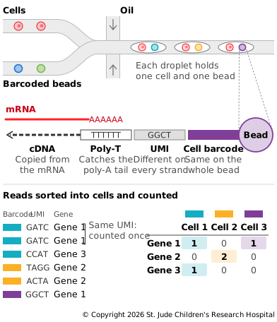
One run can capture thousands to tens of thousands of cells. Not every cell is captured, and a small share of droplets catch two cells at once, called doublets, which software tries to find and remove.
Depth and Read Setup
Depth is counted per cell, and it's low: a typical experiment reads far fewer molecules from each cell than bulk RNA-seq reads from a whole sample. Most methods sequence only one end of each mRNA, enough to tell which gene it came from and count it, but not to see its whole sequence. One read of each pair carries the barcode and UMI, and the other carries the gene sequence.
Tradeoffs
Pros
- Cell types. Single-cell RNA-seq finds the kinds of cells in a sample, including rare ones, by grouping cells with similar expression.
- Changes along a path. It can show how cells change along a path, such as a stem cell maturing.
- Where a gene is expressed. It shows which cell types express a gene of interest.
- Tumor and normal cells. In cancer, it can separate tumor cells from the normal cells around them.
Cons
- Each cell gives little data. Only a fraction of each cell's mRNA is captured, so many genes have zero reads in a given cell. Most of those zeros reflect how few molecules were sampled, not a gene that's truly off (Svensson, 2020).
- Breaking apart the tissue changes it. Separating cells can stress them, which switches on stress genes before the cells are captured (van den Brink et al., 2017). It also loses where each cell was in the tissue. Sequencing single nuclei instead of whole cells avoids some of these problems and works on frozen tissue (Habib et al., 2017).
- Cost per cell. Thousands of cells per sample add up, so experiments often include fewer samples than bulk studies.
Example: A New Cell Type for Cystic Fibrosis
In 2018, two groups used single-cell RNA-seq to map the cells lining the airways of mice and people (Montoro et al., 2018; Plasschaert et al., 2018). Both found a rare cell type no one had described before, which they called the pulmonary ionocyte. Ionocytes make up only about 1% of the cells in the airway lining, but they express CFTR, the gene behind cystic fibrosis, at much higher levels than the cells around them. In mice, they were the source of most of the airway's CFTR RNA.
Bulk RNA-seq averages CFTR expression across all the airway's cells, so there was no way to see that so much of it came from one rare cell type. Seeing each cell separately changed which cells researchers study to understand the disease.
Up Next
This chapter followed a sample from collection to a set of reads, and showed how the choice of assay decides what those reads can tell you. The next chapter, From Reads to Variants, follows the reads from there, starting with mapping them to the reference.