Signaling Pathways
A cell in the body doesn't decide to divide on its own. It waits for signals from outside, such as growth factors, small proteins released by other cells. The signal arrives at the cell's surface, but the genes that carry out division sit in the nucleus, so a chain of proteins has to carry the message inward. Each protein in the chain switches on the next, like runners passing a baton. A chain like this is called a signaling pathway.
More than 600 genes are now known to drive cancer when they're mutated (IntOGen), but many of their proteins work in the same few pathways. In a study of more than 9,000 tumors from 33 kinds of cancer, 89% had at least one driver change in just ten well-known pathways (Sanchez-Vega et al., 2018). This page follows one relay that is switched on in a large share of cancers, then two brakes, RB and p53, that a tumor has to get past.
Passing the Signal
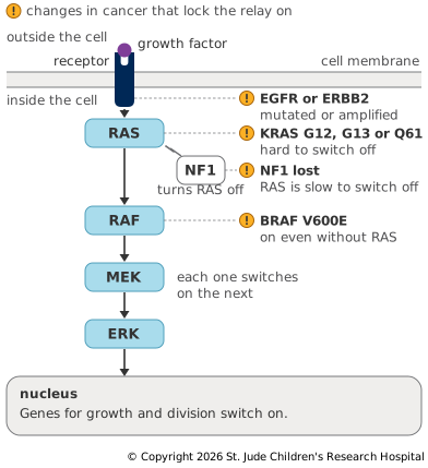
The best-studied relay is the RAS–MAPK pathway, which passes growth signals from the cell surface to the nucleus. It starts when a growth factor binds a receptor, a protein that spans the cell membrane with one end outside the cell and the other inside. One example is EGFR, the receptor for epidermal growth factor. EGFR is a kinase, an enzyme that adds phosphate groups to proteins, and Kinases as Switches shows how a phosphate can switch a protein on or off. When the growth factor binds, the receptor switches on and draws in helper proteins that switch on RAS.
RAS works like a light switch. It holds a small molecule called GDP when it is off and swaps it for a similar molecule, GTP, to switch on. RAS can turn GTP back into GDP and switch itself off, but only slowly. Proteins called GAPs speed this up, so a pulse of signal fades quickly once the growth factor is gone (Simanshu et al., 2017).
While RAS is on, it draws in a kinase called RAF. RAF adds a phosphate to a second kinase, MEK, and MEK does the same to a third, ERK. ERK adds phosphates to many proteins, which together control whether the cell grows, divides, survives, moves or matures (Lavoie et al., 2020). Among the genes that growth signals switch on is the one for cyclin D1, which comes back in the RB brake below (Asghar et al., 2015).
Locking the Relay On
A change in any of several proteins in the relay can keep the signal going after it should have faded (Figure 2.6). Mutations in RAS genes drive about a third of all human cancers (National Cancer Institute). The changes at amino acids 12, 13 and 61 of KRAS that stand out in Figure 2.4 make RAS resistant to GAPs. Once it picks up GTP, it switches itself off only slowly, so it spends far more of its time switched on.
BRAF, one of the genes for RAF, sits one step further down. When researchers screened cancers for BRAF mutations in 2002, they found them in 66% of the melanomas they tested, and a single change, now called V600E, made up 80% of them. The changed protein was a more active kinase, and cancer cells that carried it kept growing without needing RAS (Davies et al., 2002).
The relay can also jam when an off switch is lost. NF1 makes neurofibromin, a GAP for RAS. In tumors that lose it, RAS stays on for longer, with an effect much like a mutation in RAS itself. NF1 is a tumor suppressor that works inside a relay of oncogenes.
Because so many genes feed into the same relay, the pathway often tells you more than any one gene. In a study of 230 lung adenocarcinomas, 33% of tumors had a KRAS mutation and 14% had an EGFR mutation, but the two were not found in the same tumor. Changes in NF1, ERBB2 and a few other genes in the relay were more common in the tumors that had neither. Altogether, 76% of the tumors had the relay switched on in some way (Cancer Genome Atlas Research Network, 2014). A tumor without a KRAS mutation may well have the same relay switched on through a different gene.
The RB Brake
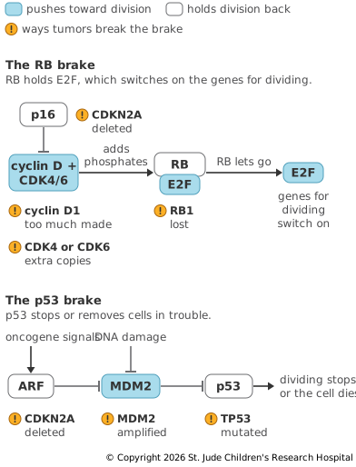
The decision to divide is made in the nucleus, where RB, the protein made by the retinoblastoma gene RB1 from Two Hits, guards it. RB holds on to proteins called E2F, which switch on the genes a cell needs to copy its DNA and divide (Dyson, 2016). Growth signals raise the level of cyclin D, which partners with the kinases CDK4 and CDK6. Together they add phosphates to RB, RB lets go of E2F and the cell commits to dividing. A protein called p16, made by the gene CDKN2A, blocks CDK4 and CDK6 and keeps the brake on (Figure 2.7).
The brake also responds to the relay. When a relay is jammed on, the cell can make more p16, which holds RB in place and halts division in a lasting state called senescence (Serrano et al., 1997). To keep growing, a tumor has to get past this, for example by losing p16.
A tumor can break the brake at any step. It can lose RB1, make too much cyclin D1, carry extra copies of CDK4 or CDK6 or delete CDKN2A. These changes tend not to appear together, and different kinds of tumor favor different ones. RB loss is a hallmark of small cell lung cancer (George et al., 2015), too much cyclin D1 is common in breast cancer (Tobin and Bergh, 2012) and losing p16 is particularly common in glioblastoma (Brennan et al., 2013). Some viruses that cause cancer make proteins that switch RB off.
The p53 Brake
The protein p53, made by TP53, responds to trouble rather than to growth signals. Most of the time a protein called MDM2 keeps the level of p53 low by marking it for destruction. Damaged DNA, or the stress caused by an oncogene, frees p53 from MDM2. DNA damage adds a phosphate to p53 that blocks MDM2 (Shieh et al., 1997), and signals from an oncogene switch on a protein called ARF (Zindy et al., 1998), which blocks MDM2 directly (Pomerantz et al., 1998). Once free, p53 switches on genes that stop the cell from dividing, such as the gene for p21, which blocks the kinases that drive the cell cycle. If the damage is too great, p53 can make the cell die instead (Kastenhuber and Lowe, 2017), as the lower half of Figure 2.7 shows. This is the checkpoint described in Genes That Guard the Genome.
TP53 is the most frequently mutated gene in human cancer (Kandoth et al., 2013), and Figure 2.5 shows where its mutations fall. A tumor can also switch off p53 without touching TP53. In a 1992 study, MDM2 was amplified in more than a third of 47 sarcomas, which suggested that the extra MDM2 helps these tumors escape p53's control (Oliner et al., 1992).
CDKN2A connects the two brakes. The same stretch of DNA is read in two different reading frames to make two unrelated proteins, p16 for the RB brake and ARF for the p53 brake (Quelle et al., 1995). Deleting the gene removes both at once. Copy Number Variation describes how tumors lose both copies of CDKN2A in many kinds of cancer.
Up Next
A tumor rarely gets all of these changes at once. How Tumors Evolve follows how they build up over time, and why tumors often find a way around the drugs aimed at them.