Protein Folding and Modification
This is a guide to genomics, so proteins only get a cameo; proteomics, the study of proteins, could fill a guide of its own. But after following a protein through transcription and translation, it felt rude to abandon it as a floppy chain at the ribosome. So here's the rest of its story, in brief.
From Chain to Shape
The ribosome releases a protein as a long chain of amino acids, but a chain alone can't do much. To work, it must fold into a precise three-dimensional shape, a process called protein folding. Some amino acids are attracted to water and others repel it, some carry an electric charge, and some can bond to each other. As the chain folds, the water-repelling amino acids tend to tuck inside and the charged ones stay on the surface, until the protein settles into a stable shape. Folding often begins while the chain is still emerging from the ribosome, and helper proteins called chaperones guide many proteins into the right shape and keep them from clumping together.
A protein's shape decides what it does. An enzyme's shape forms a pocket that fits only the molecules it acts on, a receptor's shape lets it recognize one particular signal, and a structural protein such as collagen forms long, strong cables. Larger proteins are often built from several domains, compact sections that each fold on their own and do one job, such as gripping DNA. Knowing which domain a variant falls in is a strong clue to what it might break. Because the amino acid sequence largely determines the shape, scientists can now predict a protein's structure from its sequence alone with programs such as AlphaFold, work recognized with the 2024 Nobel Prize in Chemistry.
This also means a small change in the DNA can ruin a protein. The most common variant behind cystic fibrosis deletes three bases from the CFTR gene. Three bases make exactly one codon, so the reading frame is kept and only one amino acid is lost (see Reading Frames). Yet that single missing amino acid is enough to make the protein misfold, and the cell destroys it before it reaches its place on the cell surface. Misfolded proteins that build up instead of being cleared are also linked to diseases such as Alzheimer's and Parkinson's.
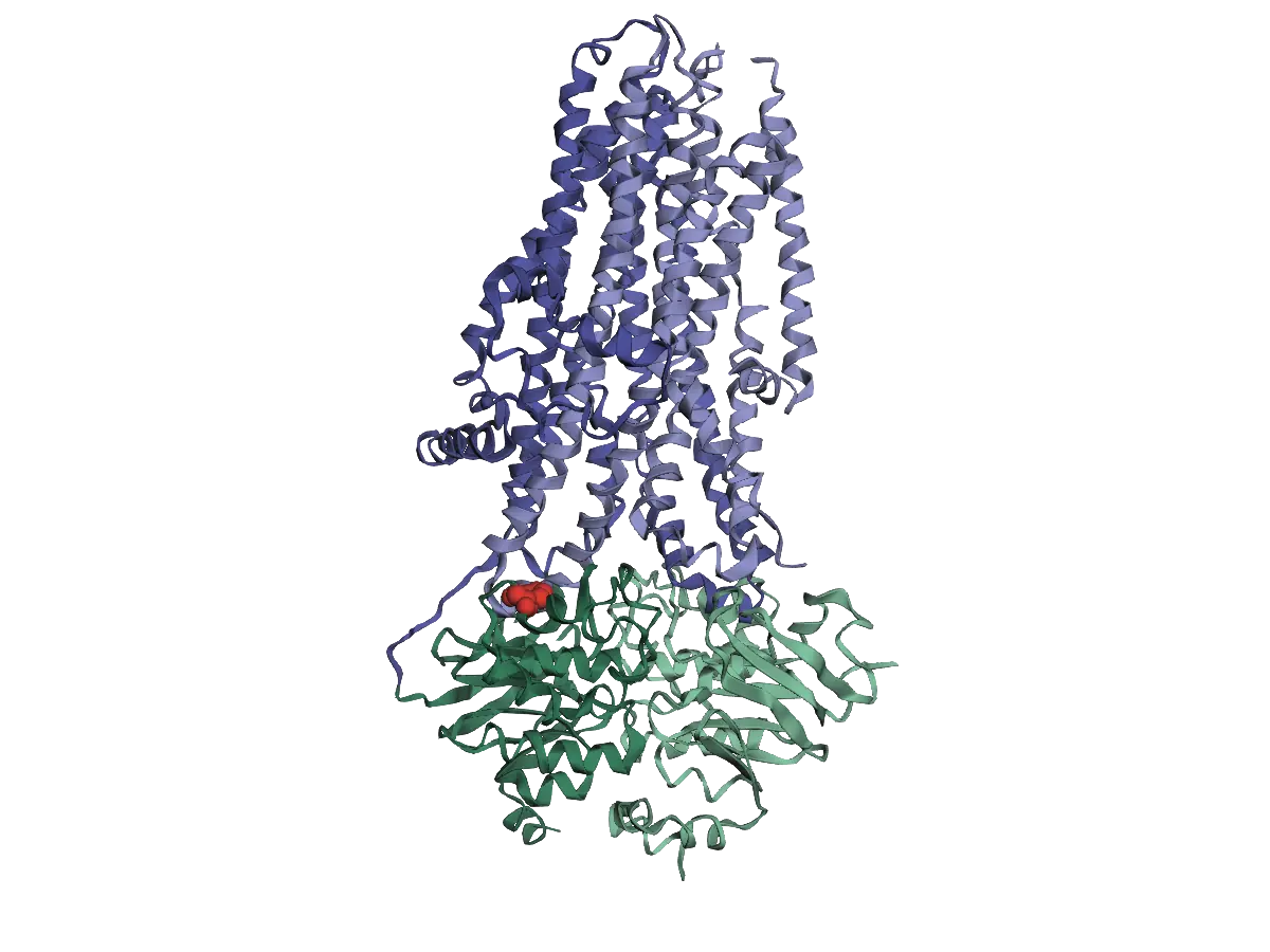
An interactive 3D model of the CFTR protein, drawn as ribbons. The two membrane domains, in two shades of blue, stand side by side at the top. The two ATP-binding domains, in two shades of green, sit below them. A red sphere marks Phe508 on the first ATP-binding domain, where it meets the membrane domains.
The CFTR protein in 3D. The two membrane domains (blue) form a channel through the cell membrane, and the two ATP-binding domains (green) sit just inside the cell. Phe508 (red) is the amino acid lost in the most common cystic fibrosis variant. Structure from PDB 6MSM (Zhang, Liu and Chen, 2018).
Getting to the Right Place
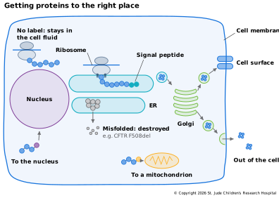
A protein also has to end up where it's needed. Proteins that work in the fluid inside the cell are simply released there, but others carry an address label. A short stretch at the start of the chain, called a signal peptide, sends the ribosome to dock on the endoplasmic reticulum (ER), a network of membranes next to the nucleus. The protein is threaded into the ER as it is made, then passed to the Golgi apparatus, which sorts it and ships it to the cell surface or out of the cell entirely. Other labels send proteins into the nucleus or the mitochondria. The ER also checks the folding of everything that passes through it, which is where the misfolded cystic fibrosis protein is caught and destroyed.
Post-Translational Modifications
Many proteins are changed further after they are made, through post-translational modifications (PTMs). Other enzymes attach small chemical groups, add sugars, or cut the chain. Hundreds of kinds are known; a few common ones are:
- Phosphorylation adds a phosphate group, which can switch a protein on or off. Enzymes called kinases add phosphates and others remove them, so the switch can flip within seconds. Kinases as Switches below looks at this switch more closely.
- Glycosylation adds chains of sugars as proteins pass through the ER and Golgi. Most of these proteins are bound for the cell surface or for export, and the sugars help them fold, stay stable and be recognized. Your ABO blood type depends on which sugars an enzyme encoded by the ABO gene adds.
- Cleavage cuts a protein into its working form. Insulin, for example, is made as one longer chain; a middle piece is cut out, leaving two shorter chains joined together.
- Ubiquitination attaches a small protein called ubiquitin, which often marks a protein for destruction.
Modifications mean that one gene can give rise to many forms of a protein, each with different activity, and that the amount of a protein in a cell doesn't tell you how much of it is active. They are also why some medicines can't be made in bacteria: bacteria can't add human-style sugars, so proteins such as therapeutic antibodies are made in mammalian cells instead.
Kinases as Switches
Phosphorylation controls so much of what a cell does, and comes up so often in cancer, that it's worth a closer look. A protein kinase, or kinase for short, is an enzyme that takes a phosphate group from ATP, the molecule cells use to carry energy, and attaches it to a serine, threonine or tyrosine in a protein, leaving ADP behind (Ardito et al., 2017). The phosphate carries a negative charge, so adding one can change the protein's shape and, with it, what the protein does. Enzymes called phosphatases take the phosphates off again, which makes the switch reversible. Human cells make about 535 protein kinases, though about 50 of them have lost the ability to add phosphates (Wilson et al., 2018).
Kinases are proteins themselves, and most of them share the same basic shape (Figure 1.13). The chain folds into two lobes, a smaller upper lobe and a larger lower lobe, joined by a short hinge. ATP fits into the cleft between the lobes, and the target protein binds right beside it (Modi & Dunbrack, 2019). A flexible stretch of the lower lobe, the activation loop, decides whether the target can get in. In many kinases that are off, the loop folds onto the surface and covers the spot where the target would bind. In a kinase called ERK, a tyrosine on the loop sits right in that spot (Zhang et al., 1994).
Many kinases are switched on when phosphates are added to their activation loop, often by another kinase. When ERK is switched on, it carries phosphates on a threonine and a tyrosine in the loop. Their negative charges pair with positively charged arginines on the kinase, and the loop refolds into a new shape that opens the site where the target binds (Canagarajah et al., 1997). Not every kinase is controlled this way, but the shape of the activation loop governs the activity of many of them (Nolen et al., 2004).
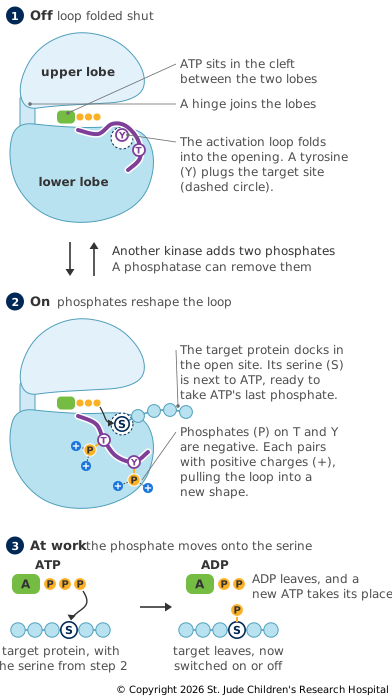
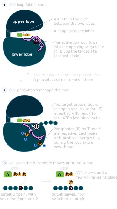
Once the phosphate has moved, the target lets go, and its new phosphate switches it on or off. ADP leaves the cleft too, a fresh ATP takes its place, and the kinase is ready for another target.
Because an active kinase can switch on another kinase, kinases often work in chains, each one passing the signal to the next. In one well-studied chain, RAF switches on MEK, and MEK switches on ERK by adding those two phosphates to its activation loop. ERK in turn adds phosphates to proteins such as ELK1 that switch on genes in the nucleus (Roskoski, 2012). Phosphatases can switch each step back off, so the signal lasts only as long as something keeps it going.
A kinase that can't be switched off keeps passing its signal on, and many cancers depend on one. A change called V600E in BRAF, a member of the RAF family, is found in many melanomas (Davies et al., 2002). It breaks up the contacts that hold BRAF's activation loop in its off position, so the kinase stays on (Wan et al., 2004). Kinases are also drug targets. The US Food and Drug Administration has approved 94 small-molecule drugs that block protein kinases, about 80 of them for cancers (Roskoski, 2026). Signaling Pathways follows this chain of kinases from the cell surface to the nucleus.
Proteins at Work
Folded, modified and delivered, a protein finally gets to work: building structures, speeding up chemical reactions, carrying signals and switching genes on and off. Added up across the trillions of cells in a body, that work produces the traits we can observe, which is where the next page picks up.