Short-Read Sequencing
The most widely used sequencers are made by Illumina. They read a few hundred bases from each fragment, which is why their data is called short-read sequencing, and they make up for the short reads with sheer numbers: a single run of a large instrument reads billions of fragments at once.
Sequencing by Synthesis
Illumina instruments use a method called sequencing by synthesis. Rather than reading a strand of DNA directly, they watch it being copied one base at a time. Illumina's own animation walks through the whole process in about five minutes. It shows the original version of the chemistry, with four dye colors. Newer instruments use fewer dyes, but the steps are the same.
Nothing loads from YouTube until you press play. Watch on YouTube
Illumina's animation of sequencing by synthesis, from library to aligned reads.
In outline:
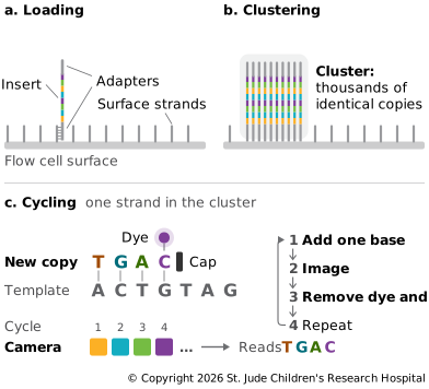
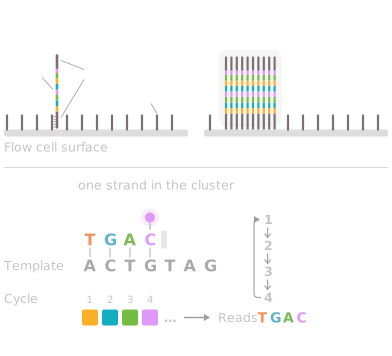
- Loading. The library is washed over a flow cell, a glass slide with narrow channels whose inner surface is coated with short DNA strands that match the adapters. Fragments stick where their adapters meet a matching strand.
- Clustering. Each fragment is copied in place, over and over, into a tight cluster of identical copies about one micron across (Bentley et al., 2008). A single molecule is too faint to see, but a cluster of copies glows brightly enough for a camera.
- Cycling. In each cycle, the instrument adds all four nucleotides, each carrying a fluorescent dye and a removable cap that stops the copying after one base. Every cluster adds exactly one base, the camera records the color at each cluster, and the dye and cap are washed off before the next cycle. Cycle 1 reads the first base of every fragment, cycle 2 the second, and so on, so 150 cycles give 150-base reads.
Software turns the images into bases, a step called base calling, and gives each base a score for how sure it is.
Quality Scores
Every base comes with a quality score, written on the Phred scale (Ewing and Green, 1998). The score Q stands for the estimated chance that the base is wrong, P, through Q = −10 × log₁₀(P). Each step of 10 is a tenfold change:
| Quality score | Chance the base is wrong | Accuracy |
|---|---|---|
| Q10 | 1 in 10 | 90% |
| Q20 | 1 in 100 | 99% |
| Q30 | 1 in 1,000 | 99.9% |
| Q40 | 1 in 10,000 | 99.99% |
Q30 is the usual yardstick: instrument specifications and run reports quote the share of bases at Q30 or above. The scores are stored next to the bases in FASTQ files, and later steps use them to decide how much to trust each base.
Quality falls along the read. In every cycle, a few molecules in a cluster fail to add a base, or add two, and fall out of step with the rest. The errors add up, so by later cycles each cluster's signal is a blur of neighboring positions. This is the main reason short reads stay short.
Paired-End Reads
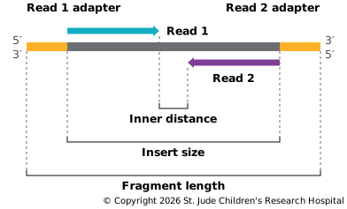
Most runs read each fragment from both ends. The instrument first reads a set number of bases from one end, called Read 1. It then copies the fragment so the other strand faces up and reads the same number of bases from the opposite end, called Read 2. Together they make a read pair. A common setup reads 150 bases from each end, written 2 × 150. The reads stay the same length, but the fragments don't. On a long fragment, the two reads leave an unread gap in the middle. On a shorter one, they overlap. And when the fragment is shorter than a read, each read runs off the end of the sample's DNA and into the adapter on the far side. Those adapter bases aren't part of the sample, so trimming tools remove them (Chen et al., 2018).
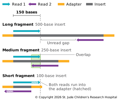
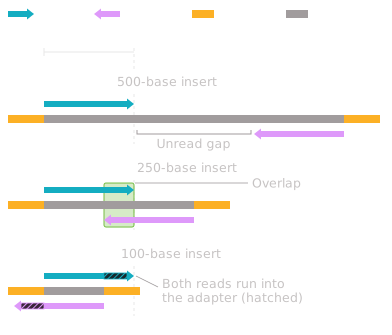
The two reads in a pair come from the same fragment, so they lie a known distance apart and face each other. The length of the fragment between the adapters is the insert size, and the unread stretch between the two reads is the inner distance.
That known spacing is useful. If one read of a pair falls in a repeat that appears in many places, its partner, in unique sequence nearby, shows which copy it came from. And when the two reads land much farther apart than the insert size allows, on different chromosomes, or facing the wrong way, they point to a rearrangement, one of the main signals in Finding Structural Variants.
Errors to Know About
Illumina reads are accurate, with roughly one error per thousand bases in a modern run (Wenger et al., 2019), but the errors aren't random, and each kind leaves its own mark:
- Mostly substitutions. Errors are almost always one base read as another. Insertions and deletions are rare.
- Worse toward the end. Error rates climb along the read, as described above, and Read 2 is usually a little worse than Read 1.
- GC bias. Regions very rich or very poor in G and C get fewer reads, partly from PCR in library preparation.
- Poly-G tails. Two-color instruments, such as the NextSeq and NovaSeq, record G as the absence of light. When a cluster's signal fades near the end of a read, the instrument sees darkness and calls a run of Gs that isn't there (Chen et al., 2018). Trimming tools remove them.
- Index hopping. On some instruments, a small share of reads carry another sample's index, as described in Adding Adapters and Indexes.
From Images to Files
During a run, the instrument writes its base calls to BCL files, one set per cycle. When the run ends, software converts them into FASTQ files, sorting reads into samples by their index on the way. A paired-end run gives each sample two FASTQ files, one for Read 1 and one for Read 2, with the reads in the same order in both. Those FASTQ files are usually where a computational team's work begins.
Other Short-Read Instruments
Illumina isn't the only maker, and the market has grown more crowded. Most of these instruments also read by synthesis, each with its own chemistry, and their output looks much the same: short reads, quality scores, FASTQ files. This list is not complete, and it changes quickly. Each company's site has more on its instruments.
| Company | Instruments | How it differs |
|---|---|---|
| Element Biosciences | AVITI | Reads with a different chemistry, called avidity sequencing |
| MGI and Complete Genomics | DNBSEQ | Copies each fragment into a ball of DNA, a DNA nanoball, instead of a cluster |
| Ultima Genomics | UG 100, UG 200 | Spreads fragments on a spinning silicon wafer to lower cost |
| Thermo Fisher (Ion Torrent) | Ion GeneStudio, Genexus | Detects the tiny change in acidity as each base is added, instead of light |