Oncogenes and Tumor Suppressors
The hallmarks come from changes in genes. Many genes help decide when a cell grows and divides, and a car is a helpful way to picture how they work together. A driver keeps a car under control with two pedals, pressing the accelerator to speed up and the brake to slow down. A cell controls its growth in a similar way. Some genes act like the accelerator, pushing the cell to grow and divide, and others act like the brakes, holding it back. Cancer can start when the accelerator sticks to the floor or when the brakes fail. A third group of genes is more like the mechanic who keeps the car in good shape: losing them doesn't make the car go faster, but faults build up more quickly, including faults in the accelerator and the brakes.
These three groups are called proto-oncogenes, tumor suppressor genes and DNA repair genes. The genetic changes that drive cancer tend to affect them (National Cancer Institute). This page covers each kind of gene, the two-hit model that explains how tumor suppressors are lost and how researchers tell the changes that drive a tumor from the ones that only come along for the ride.
Oncogenes
Proto-oncogenes are the accelerator, normal genes that help cells grow and divide. When one is changed so that its protein is more active than normal, or the cell makes too much of it, it becomes an oncogene, and the cell can grow and survive when it shouldn't (National Cancer Institute). The idea came from a virus that causes cancer in birds. Its cancer-causing gene, src, turned out to be a changed copy of a normal gene that the virus had picked up from a cell it infected (Berger et al., 2011).
Proto-oncogenes become oncogenes in three main ways (Berger et al., 2011; Vogelstein et al., 2013). The Genomic Variation chapter shows an example of each.
- A point mutation changes the protein so that it stays switched on. BRAF V600E, in Single Nucleotide Variation, works this way.
- Amplification gives the cell many extra copies of the gene, often 10 to 100, so it makes far more of the protein. ERBB2 in breast cancer is an example, in Copy Number Variation.
- A chromosome rearrangement can join part of the gene to part of another gene, making a new fusion protein, or move the gene next to DNA that switches it on too strongly. In BCR-ABL1, a fusion keeps a kinase permanently switched on.
Because the changed protein pushes the cell to grow whatever the other copy of the gene is doing, a change in one of the two copies is usually enough. Oncogenes are said to act dominantly, and the change is a gain of function (Lee and Muller, 2010).
Tumor Suppressors
Tumor suppressor genes are the brakes. Like proto-oncogenes, they help control cell growth and division (National Cancer Institute), but they hold it back. A tumor gains from them when they stop working, so the changes that help a tumor cause a loss of function. For most tumor suppressors, one working copy is enough to do the job, so a cell has to lose both copies before it escapes their control. Inside the cell, tumor suppressors act recessively (Carbone and Minna, 1993).
There are many ways to lose a copy (Berger et al., 2011):
- a nonsense or frameshift mutation that cuts the protein short;
- a deletion of the gene, as when a tumor loses both copies of CDKN2A (Copy Number Variation);
- copy-neutral loss of heterozygosity, where the cell loses its working copy and duplicates the broken one;
- chemical marks on the gene's promoter that switch it off without changing its sequence. In most colorectal cancers with microsatellite instability that were not inherited, the DNA repair gene MLH1 is switched off this way (Herman et al., 1998).
Not every tumor suppressor follows the two-copy rule. For some, such as TP53 and PTEN, losing one copy, or even making a little less of the protein, can be enough to help a tumor (Berger et al., 2011).
Two Hits
The need to lose both copies solved a puzzle about inherited cancer. In families that carry a broken copy of a tumor suppressor, the risk of cancer is passed down as a dominant trait. Inside the cell, though, the gene acts recessively (Berger et al., 2011).
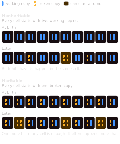
In 1971, Alfred Knudson worked out why by studying retinoblastoma, an eye cancer of young children. He proposed that the tumor needs two hits (Knudson, 1971). In the heritable form, a child is born with the first hit in every cell, so one more hit in any cell of the developing retina is enough. In the nonheritable form, both hits have to happen in the same cell (Figure 2.3). A second hit somewhere in the retina is so likely that almost every child born with the first hit develops retinoblastoma (Berger et al., 2011). These children have an average of three tumors, which explains why some have tumors in both eyes or several in one eye (Knudson, 1971). They are also diagnosed at a younger age (Berger et al., 2011). A tumor in both eyes, or several in one eye, suggests the heritable form (National Cancer Institute).
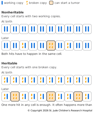
The gene was later found to be RB1. When researchers compared tumors with normal cells from the same children, the tumor cells had lost the working copy and kept only the broken one, often through losing a chromosome or through recombination (Cavenee et al., 1983). When the remaining chromosome is copied, or the change comes from recombination, the cell still has two copies of the gene. This is the copy-neutral loss of heterozygosity that shows up in allele fractions.
A child with heritable retinoblastoma may have inherited the broken copy from a parent, or it may have arisen new in a sperm or egg cell or early in development (National Cancer Institute). Either way, the two-hit model explains why inheriting one broken copy of an important gene can create a predisposition to cancer without causing it directly. Inherited Risk returns to this idea.
Genes That Guard the Genome
DNA repair genes fix damaged DNA. Cells with mutations in these genes tend to pick up more mutations and chromosome changes (National Cancer Institute). Losing them doesn't make a cell grow on its own. It speeds up the changes in other genes that do.
- Mismatch repair genes, such as MLH1 and MSH2, fix mistakes made when DNA is copied. Tumors that have lost mismatch repair can carry thousands of mutations, and the microsatellite instability they cause shows up clearly in sequencing data.
- Checkpoint genes, such as TP53 and ATM, help the body get rid of cells whose DNA is damaged. When they are lost, damaged cells survive and keep dividing, including cells that carry large changes, such as a translocation or an extra chromosome.
Inherited Risk covers what happens when a child is born with a broken copy of one of these genes.
Drivers and Passengers
Only a few of a tumor's mutations help it grow. In common solid tumors, such as those of the colon, breast, brain and pancreas, an average of 33 to 66 genes carry mutations that would be expected to change their proteins. A typical tumor has only two to eight driver mutations, and the rest are passengers. Single Nucleotide Variation shows how a mutation that recurs across many tumors points to a driver.
Where the mutations fall in a gene also shows which kind of driver it is. Oncogenes are mutated again and again at the same few positions, because only certain changes switch the protein on. Tumor suppressors are often hit by changes that cut the protein short, spread along much of the gene, because a break in many different places can switch it off. Vogelstein and colleagues turned this into the 20/20 rule. A gene counts as an oncogene if more than 20% of its recorded mutations are missense changes at recurring positions, and as a tumor suppressor if more than 20% inactivate it. In 2013, 125 genes met the rule, 54 oncogenes and 71 tumor suppressors.
Figure 2.4 shows the oncogene pattern in real tumors. It plots the mutations in KRAS found in tumors sequenced for the NCI Genomic Data Commons, drawn the same way as the BRAF plot in Figure 3.4. The bar along the bottom is the KRAS protein, from its first amino acid on the left to its last on the right. Each circle above it is one protein change, and the number inside is how many tumors carry it. The labels follow the same naming as BRAF V600E, so G12D means that the glycine (G) at position 12 has become an aspartic acid (D).
Most of the large circles sit at just three positions, amino acids 12, 13 and 61, and the most common changes there are each found in hundreds of tumors. These are the positions where changes switch on KRAS and the two closely related RAS genes, and KRAS is the most often mutated of the three (Prior et al., 2012). The rest of the protein carries smaller circles, most of them found in only a few tumors.
Loading KRAS variants from ProteinPaint…
A lollipop plot of the KRAS protein, about 190 amino acids long, drawn as a bar with its domains shaded. Somatic variants from tumors stand above it as circles on stems, sized and numbered by how many tumors carry each one. Tall stacks of large circles sit at positions 12, 13 and 61, and the largest, G12D, G12V and G12C, are each found in hundreds of tumors. Small circles, each found in only a few tumors, are scattered along the rest of the protein.
Somatic variants in KRAS from tumors in the NCI Genomic Data Commons. Each circle is one protein change, labeled with its name and sized by the number of tumors that carry it. Most of the recurrent changes fall at amino acids 12, 13 and 61. Drawn with ProteinPaint (Zhou et al., 2016), which loads current data, so the counts grow as new tumors are added.
The pattern can overturn a first guess. IDH1 was first thought to be a tumor suppressor, but nearly all of its mutations change the same amino acid, at position 132, which marked it as an oncogene.
The pattern is a strong clue rather than a rule, and TP53 shows why (Figure 2.5). It is a tumor suppressor, but about 90% of its mutations are missense changes. They fall at about 190 positions in the part of the protein that binds DNA, and just eight changes, at six of those positions, account for about 28% of all its mutations. These changes weaken the protein's hold on DNA, so they switch off its normal job rather than switching it on. Some of the changed proteins may also gain new activities that help the tumor (Baugh et al., 2018).
Loading TP53 variants from ProteinPaint…
A lollipop plot of the TP53 protein, about 390 amino acids long, drawn as a bar with its domains shaded. Somatic variants from tumors stand above it as circles on stems, sized and numbered by how many tumors carry each one. Most circles are missense changes packed into the middle of the protein, the part that binds DNA, and a handful of positions there are found in far more tumors than the rest. Smaller circles for changes that cut the protein short are scattered along its length.
Somatic variants in TP53 from tumors in the NCI Genomic Data Commons. Each circle is one protein change, labeled with its name and sized by the number of tumors that carry it. Unlike most tumor suppressors, TP53 is hit mostly by missense changes, and a few positions recur in many tumors. Drawn with ProteinPaint (Zhou et al., 2016), which loads current data, so the counts grow as new tumors are added.
Up Next
Driver genes don't act alone. Their proteins work in a small number of pathways that control whether a cell divides, whether it survives and how it maintains its genome. Signaling Pathways follows some of the most important.