Assay for Transposase-Accessible Chromatin Using Sequencing (ATAC-seq)
ATAC-seq (assay for transposase-accessible chromatin using sequencing) finds the parts of the genome that are open. Most DNA in the nucleus is wrapped tightly around histones, out of reach of the proteins that read genes. The stretches left open include the promoters and enhancers a cell is using, so a map of open DNA is a map of where the genome could be active.
What It Measures
ATAC-seq measures accessibility. DNA wraps around histones in units called nucleosomes, each holding about 150 bases, and the DNA between nucleosomes is easier to reach. Where chromatin is loosely packed, there are wider gaps, and those gaps are where transcription factors bind.
Unlike ChIP-seq, ATAC-seq doesn't target a particular protein. It shows every open region at once.
How the Library Is Made
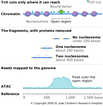
ATAC-seq uses the same enzyme as tagmentation: a transposase, Tn5, loaded with sequencing adapters. The enzyme is added to intact nuclei, where it cuts DNA and attaches adapters in the same step, but only where it can reach. Open DNA gets cut often; DNA wrapped in nucleosomes rarely does. The tagged fragments are amplified and sequenced.
The original method took two steps and as few as 500 to 50,000 cells (Buenrostro et al., 2013), much less than earlier ways of mapping open chromatin. One problem is that the DNA in mitochondria isn't packed into nucleosomes, so the enzyme cuts it freely, and many reads can be wasted on it. An improved protocol, Omni-ATAC, cut the number of reads from mitochondrial DNA 13-fold (Corces et al., 2017).
Depth and Read Setup
Reads are mapped, and regions where they pile up are called peaks, just as in ChIP-seq. ATAC-seq is read paired-end, because the two reads of a pair give each fragment's exact length, and fragment length shows what lay between the two cuts. The enzyme can't cut DNA that is wrapped around a nucleosome, only the DNA on either side of it:
- Under about 100 bases. Both cuts landed in the same stretch of open DNA, with no nucleosome between them.
- About 200 bases. The cuts landed on either side of one nucleosome, so the fragment holds that nucleosome's DNA plus a little open DNA at each end.
- About 400 or 600 bases. The fragment spans two or three nucleosomes in a row.
Sorting fragments by length this way shows where individual nucleosomes sit inside an open region, not just that the region is open (Buenrostro et al., 2013).
Tradeoffs
Pros
- The whole genome at once. ATAC-seq finds open regions across the genome.
- Fewer cells, less work. It needs fewer cells and less work than other methods.
- Footprints. Where a protein is bound inside an open region, it shields the DNA beneath it from the enzyme, leaving a small gap in the cuts called a footprint. Footprints at the binding sequence of a protein called CTCF were visible in the original study.
Cons
What it can't show is which protein is bound, or whether an open region is actually doing anything. Open isn't the same as active. ATAC-seq is often paired with RNA-seq or ChIP-seq to connect open regions to the genes they control.
Example: A Blood Sample in an Afternoon
The first ATAC-seq paper showed how quick the method could be. Starting from a blood sample, the authors had a finished sequencing library in 275 minutes (Buenrostro et al., 2013). They mapped open chromatin in one person's T cells, a kind of immune cell, on consecutive days, showing that a person's chromatin could be profiled within a day. Since then, ATAC-seq has become one of the most widely used ways to map open chromatin, including in single cells.